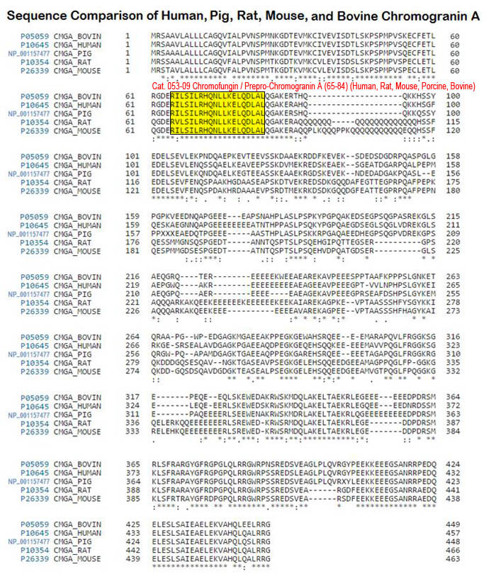
Endogenous chromogranin A (CgA)-derived peptides are secreted by nervous, endocrine and immune cells. Chromofungin (Chr: CgA47-66) is one of these peptides that display antimicrobial activities and activate neutrophils, with important implications in inflammation and innate immunity. The aim of the present study is to examine the effects of Chr on isolated and Langendorff perfused rat hearts. The study was performed by using the isolated and Langendorff perfused rat hearts, Elisa assay and real-time PCR. We found that, under basal conditions, increasing doses (11-165nM) of Chr induced negative inotropic effects without changing coronary pressure. This action was mediated by the AKT/eNOS/cGMP/PKG pathway. We also found that Chr acted as a postconditioning (PostC) agent against ischemia/reperfusion (I/R) damages, reducing infarct size and LDH level. Cardioprotection involved PI3K, RISK pathway, MitoKATP and miRNA-21. We suggest that Chr directly affects heart performance, protects against I/R myocardial injuries through the activation of prosurvival kinases. Results may propose Chr as a new physiological neuroendocrine modulator able to prevent heart dysfunctions, also encouraging the clarification of its clinical potential.
Filice E, Pasqua T, Quintieri AM et al., Peptides. 2015 Jul 4;71:40-48. doi: 10.1016/j.peptides.2015.06.013. [Epub ahead of print]
BACKGROUND: Antimicrobial peptides derived from the natural processing of chromogranin A (CgA) are co-secreted with catecholamines upon stimulation of chromaffin cells. Since PMNs play a central role in innate immunity, we examine responses by PMNs following stimulation by two antimicrobial CgA-derived peptides.
METHODOLOGY/PRINCIPAL FINDINGS: PMNs were treated with different concentrations of CgA-derived peptides in presence of several drugs. Calcium mobilization was observed by using flow cytometry and calcium imaging experiments. Immunocytochemistry and confocal microscopy have shown the intracellular localization of the peptides. The calmodulin-binding and iPLA2 activating properties of the peptides were shown by Surface Plasmon Resonance and iPLA2 activity assays. Finally, a proteomic analysis of the material released after PMNs treatment with CgA-derived peptides was performed by using HPLC and Nano-LC MS-MS. By using flow cytometry we first observed that after 15 s, in presence of extracellular calcium, Chromofungin (CHR) or Catestatin (CAT) induce a concentration-dependent transient increase of intracellular calcium. In contrast, in absence of extra cellular calcium the peptides are unable to induce calcium depletion from the stores after 10 minutes exposure. Treatment with 2-APB (2-aminoethoxydiphenyl borate), a store operated channels (SOCs) blocker, inhibits completely the calcium entry, as shown by calcium imaging. We also showed that they activate iPLA2 as the two CaM-binding factors (W7 and CMZ) and that the two sequences can be aligned with the two CaM-binding domains reported for iPLA2. We finally analyzed by HPLC and Nano-LC MS-MS the material released by PMNs following stimulation by CHR and CAT. We characterized several factors important for inflammation and innate immunity.
CONCLUSIONS/SIGNIFICANCE: For the first time, we demonstrate that CHR and CAT, penetrate into PMNs, inducing extracellular calcium entry by a CaM-regulated iPLA2 pathway. Our study highlights the role of two CgA-derived peptides in the active communication between neuroendocrine and immune systems.
Zhang D, Shooshtarizadeh P, Laventie BJ, et al., PLoS One. 2009;4(2):e4501. doi: 10.1371/journal.pone.0004501. Epub 2009 Feb 19.
Chromogranin A (CgA), an acidic granule protein of the regulated secretory pathway in the diffuse neuroendocrine system, is postulated to serve as a prohormone for regulatory peptides. Betagranin (rCgA(1-128)), the first N-terminal cleavage product of rat CgA, is 87% homologous to the bovine vasostatin I (bCgA(1-76)), previously shown to be vasoinhibitory in bovine resistance arteries. In this study the vasoactivity of homologous rat and bovine peptides was investigated in the rat posterior cerebral artery. Firstly, we examined the interaction of rhodamine (Rh)-labelled bCgA(7-40) and bCgA(47-70) with elements of the arterial wall by fluorescence microscopy. Secondly, rCgA(7-57), bCgA(1-40), bCgA(7-40) and bCgA(47-66) (chromofungin) were studied for effects on arterial tone and intracellular calcium as function of pressure in an arteriograph. Although without dilator or constrictor responses at 60-150 mm Hg, the rat peptide (rCgA(7-57)) evoked a significant delay in the onset of forced dilatation at 170 mm Hg, in contrast to the bovine peptides bCgA(1-40), bCgA(7-40) and bCgA(47-66) (chromofungin). Neither Rh-bCgA(7-40) nor Rh-bCgA(47-70) stained the endothelial layer, while Rh-bCgA(47-70) but not Rh-bCgA(7-40) stained the smooth muscle compartment. Analogously, bCgA(47-66) but not bCgA(7-40) reduced intracellular calcium, however without modifying the myogenic response. Thus, the betagranin peptide rCgA(7-57) and the two bovine chromofungin-containing peptides, highly homologous to the corresponding sequence (rCgA(47-66)), affected the rat cerebral artery without vasodilator effects, indicating significant species differences in vasoactivity of the N-terminal domain of CgA.
Mandalà M, Brekke JF, Serck-Hanssen G et al., Regul Pept. 2005 Jan 15;124(1-3):73-80.