|
|
| DILP 6 |
an IGF-like peptide secreted from fat and glial cells, regulate postfeeding and nonfeeding growth in Drosophila |
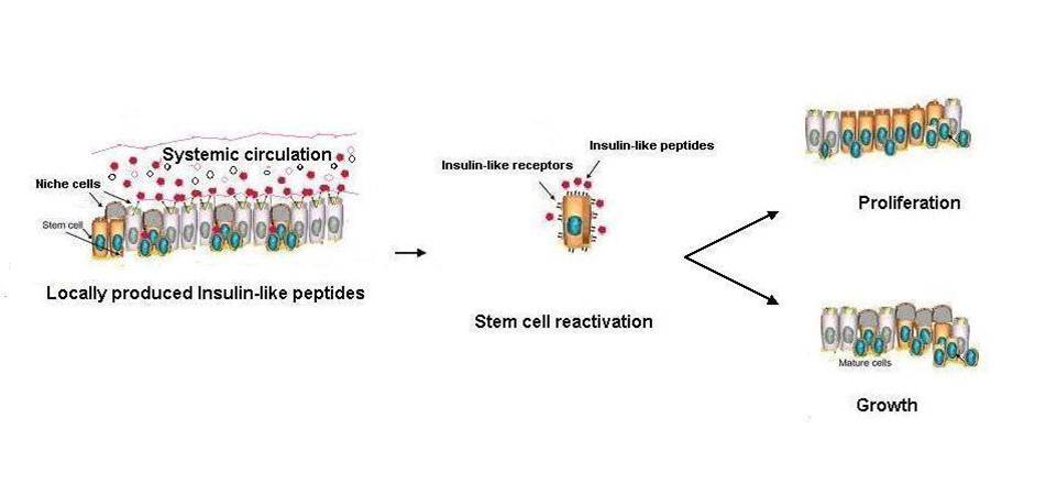
Diverse systemic signals stimulate niche cells to secrete insulin-like peptides. Such insulin-like peptides (e.g. DILP6 and DILP3) bind to cognate receptors expressed by stem cells and change their behavior, triggering growth and proliferation.
See the perspectives in “Insulin Finds its Niche.” by Seth W. Cheetham, and Andrea H. Brand, Science 340, 817-818, May 17, 2013.

|
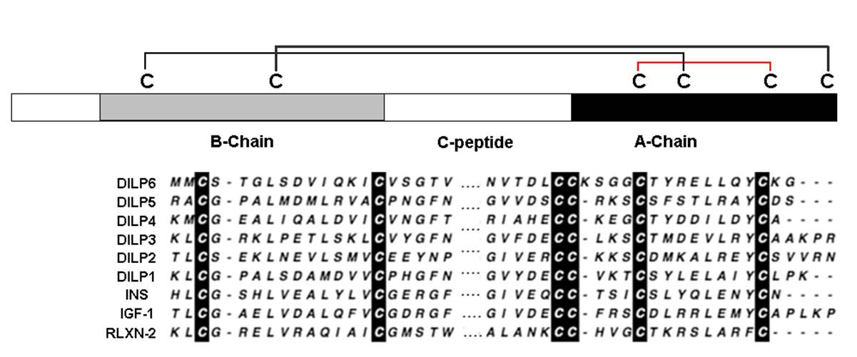
Reduced insulin/IGF signaling extends lifespan in diverse species, including Drosophila melanogaster where the genome encodes seven insulin-like peptides (dilp1-7). Of these, reduced dilp2 expressed in the brain has been associated with longevity assurance when over-expression of dfoxo in fat bodies extends lifespan. Here, we show that the insulin-regulated transcription factor dFOXO positively modulates dilp6 mRNA in adult fat body. Over-expression of dilp6 in adult fat body extends lifespan and increases longevity-associated metabolic phenotypes. Adult fat body dilp6 expression represses dilp2 and dilp5 mRNA in the brain, and the secretion of DILP2 into the hemolymph. The longevity benefit of expressing dfoxo in fat body, and the nonautonomous effect of fat body dfoxo upon brain dilp expression, is blocked by simultaneously repressing dilp6 by RNAi in fat body. dilp6 thus appears to bridge dFOXO, adipose tissue and brain endocrine function to regulate Drosophila longevity.
Bai H, Kang P, Tatar M., Aging Cell. 2012 Dec;11(6):978-85. doi: 10.1111/acel.12000. Epub 2012 Sep 18.
|
|
|
Many stem, progenitor and cancer cells undergo periods of mitotic quiescence from which they can be reactivated. The signals triggering entry into and exit from this reversible dormant state are not well understood. In the developing Drosophila central nervous system, multipotent self-renewing progenitors called neuroblasts undergo quiescence in a stereotypical spatiotemporal pattern. Entry into quiescence is regulated by Hox proteins and an internal neuroblast timer. Exit from quiescence (reactivation) is subject to a nutritional checkpoint requiring dietary amino acids. Organ co-cultures also implicate an unidentified signal from an adipose/hepatic-like tissue called the fat body. Here we provide in vivo evidence that Slimfast amino-acid sensing and Target of rapamycin (TOR) signalling activate a fat-body-derived signal (FDS) required for neuroblast reactivation. Downstream of this signal, Insulin-like receptor signalling and the Phosphatidylinositol 3-kinase (PI3K)/TOR network are required in neuroblasts for exit from quiescence. We demonstrate that nutritionally regulated glial cells provide the source of Insulin-like peptides (ILPs) relevant for timely neuroblast reactivation but not for overall larval growth. Conversely, ILPs secreted into the haemolymph by median neurosecretory cells systemically control organismal size but do not reactivate neuroblasts. Drosophila thus contains two segregated ILP pools, one regulating proliferation within the central nervous system and the other controlling tissue growth systemically. Our findings support a model in which amino acids trigger the cell cycle re-entry of neural progenitors via a fat-body-glia-neuroblasts relay. This mechanism indicates that dietary nutrients and remote organs, as well as local niches, are key regulators of transitions in stem-cell behaviour.
Sousa-Nunes R, Yee LL, Gould AP. et al, Nature. 2011 Mar 24;471(7339):508-12. doi: 10.1038/nature09867. Epub 2011 Feb 23.
|
|
|
In metazoans, tissue growth relies on the availability of nutrients--stored internally or obtained from the environment--and the resulting activation of insulin/IGF signaling (IIS). In Drosophila, growth is mediated by seven Drosophila insulin-like peptides (Dilps), acting through a canonical IIS pathway. During the larval period, animals feed and Dilps produced by the brain couple nutrient uptake with systemic growth. We show here that, during metamorphosis, when feeding stops, a specific DILP (Dilp6) is produced by the fat body and relays the growth signal. Expression of DILP6 during pupal development is controlled by the steroid hormone ecdysone. Remarkably, DILP6 expression is also induced upon starvation, and both its developmental and environmental expression require the Drosophila FoxO transcription factor. This study reveals a specific class of ILPs induced upon metabolic stress that promotes growth in conditions of nutritional deprivation or following developmentally induced cessation of feeding.
Slaidina M, Delanoue R, Gronke S, Partridge L, Léopold P. Dev Cell. 2009 Dec;17(6):874-84. doi: 10.1016/j.devcel.2009.10.009.
|
|
|
Members of the insulin family of peptides have conserved roles in the regulation of growth and metabolism in a wide variety of metazoans. Here we show that Drosophila insulin-like peptide 6 (DILP6), which is structurally similar to vertebrate insulin-like growth factor (IGF), is predominantly expressed in the fat body, a functional equivalent of the vertebrate liver and adipocytes. This expression occurs during the postfeeding stage under the direct regulation of ecdysteroid. We further reveal that dilp6 mutants show growth defects during the postfeeding stage, which results in reduced adult body size through a decrease in cell number. This phenotype is rescued by fat body-specific expression of dilp6. These data indicate that DILP6 is a functional, as well as a structural, counterpart of vertebrate IGFs. Our data provide in vivo evidence for a role of ILPs in determining adult body size through the regulation of postfeeding growth.
Okamoto N, YOkamoto N, Yamanaka N et al, Dev Cell. 2009 Dec;17(6):885-91. doi: 10.1016/j.devcel.2009.10.008.
|
|
|
|
DILP 8
%DILP%
|
|
|


