Neuropeptides are small protein-like signaling molecules with diverse roles in regulating neural functions such as sleep/wake cycles, pain modulation, synaptic plasticity, and learning and memory. Numerous drugs designed to target neuropeptides, their receptors, or relevant pathways have been developed in the past few decades. Hence, the discovery and characterization of new neuropeptides and their functions have received considerable attention from scientific research. Computational bioinformatics coupled with functional assays are powerful tools to address the difficulties in discovering new bioactive peptides. In this study, a new bioinformatic strategy was designed to screen full length human and mouse cDNA databases to search for novel peptides. One was discovered and named peptide Lv because of its ability to enhance L-type voltage-gated calcium channel (L-VGCC) currents in retinal photoreceptors. Using matrix-assisted laser desorption/ionization-time of flight mass spectrometry (MALDI-TOF MS), peptide Lv was detected in the culture media, which indicated that it was secreted from 661W cells transfected with the gene. In vitro treatments with either glutathione S-transferase (GST) fusion peptide Lv or synthesized peptide Lv enhanced L-VGCC channel activities in cone photoreceptors. At the molecular level, peptide Lv stimulated cAMP production, enhanced phosphorylation of extracellular signal-regulated kinase (ERK), and increased the protein expression of L-VGCCα1 subunits in cone photoreceptors. Therefore, the biological activities of peptide Lv may be very important in the modulation of L-VGCC dependent neural plasticity.
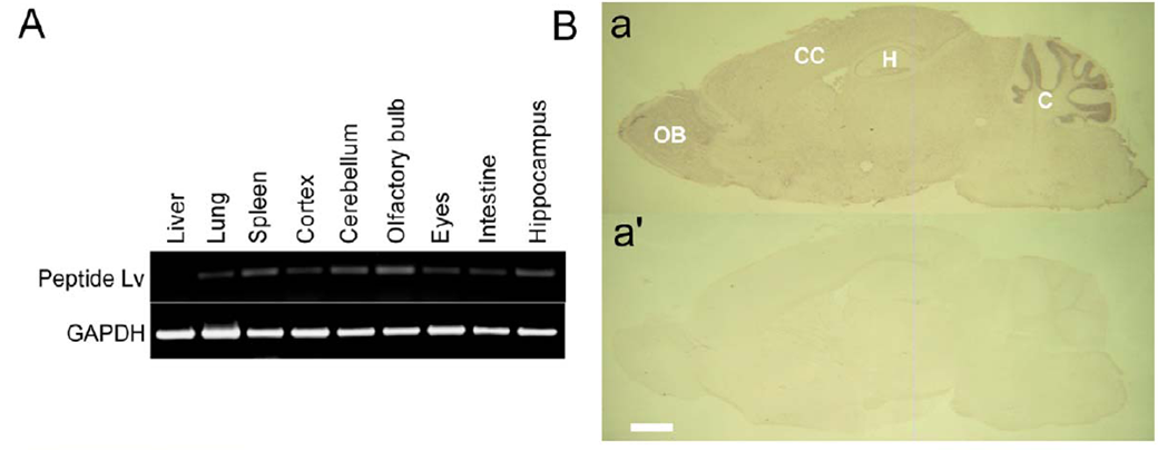
Expression of peptide Lv mRNA in mouse tissues.
(A) Gene expression of peptide Lv in various mouse tissues was determined by RTPCR. The mRNA of peptide Lv was detected in the liver, lung, spleen, intestine, eyes, cerebral cortex (cortex), cerebellum, olfactory bulb, and hippocampus, with relatively higher expression levels in the spleen, cerebellum, olfactory bulb, and hippocampus.
(B) The mRNA expression of
peptide Lv in the mouse brain and retina was detected by in situ hybridization. Adult mouse brain sagittal sections and retina sections were cut at 12 mm thickness and processed. The sense probe served as the negative control (a’). Propeptide Lv mRNA was strongly expressed in the cerebellum.
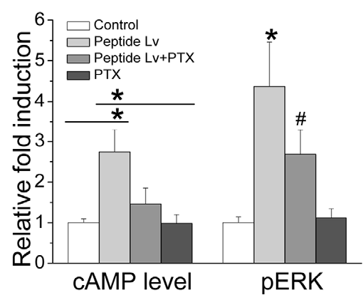
The effect of peptide Lv on pERK and cAMP is dampened by PTX. Peptide Lv increased cAMP levels (2.7460 X) and the phosphorylation of ERK (4.3761 X) after 4 hours of treatment (D). However, PTX blocked the effects of peptide Lv (n = 5 for each treatment). For cAMP levels,
*indicates that the peptide Lv treated group is significantly higher than the control and PTX treated groups (One-way ANOVA, Tukey post hoc, *p,0.05). For pERK levels, *indicates that the peptide Lv treated group is significantly higher than all other groups, while #indicates that the peptide Lv+PTX group is significantly higher than the control and PTX treated groups (One-way ANOVA, Tukey post hoc, both *and #indicate p,0.05).
Shi L et al., PLoS One. 2012;7(8):e43091. Epub 2012 Aug 13.