Vaspin
(Visceral Adipose Tissue-derived Serpin)
An Insulin-sensitizing
Adipocytokine
Visceral adipose
tissue-derived serine protease inhibitor: a unique insulin-sensitizing
adipocytokine in obesity
There is a rapid global
rise in obesity, and the link between obesity and diabetes
remains somewhat obscure. We identified an adipocytokine,
designated as visceral adipose tissue-derived serpin (vaspin),
which is a member of serine protease inhibitor family. Vaspin
cDNA was isolated by from visceral white adipose tissues (WATs)
of Otsuka Long-Evans Tokushima fatty (OLETF) rat, an animal
model of abdominal obesity with type 2 diabetes. Rat, mouse,
and human vaspins are made up of 392, 394, and 395 amino acids,
respectively; exhibit approximately 40% homology with alpha1-antitrypsin;
and are related to serine protease inhibitor family. Vaspin
was barely detectable in rats at 6 wk and was highly expressed
in adipocytes of visceral WATs at 30 wk, the age when obesity,
body weight, and insulin levels peak in OLETF rats. The tissue
expression of vaspin and its serum levels decrease with worsening
of diabetes and body weight loss at 50 wk. The expression
and serum levels were normalized with the treatment of insulin
or insulin-sensitizing agent, pioglitazone, in OLETF rats.
Administration of vaspin to obese CRL:CD-1 (ICR) (ICR) mice
fed with high-fat high-sucrose chow improved glucose tolerance
and insulin sensitivity reflected by normalized serum glucose
levels. It also led to the reversal of altered expression
of genes relevant to insulin resistance, e.g., leptin, resistin,
TNFalpha, glucose transporter-4, and adiponectin. In DNA chip
analyses, vaspin treatment resulted in the reversal of expression
in approximately 50% of the high-fat high-sucrose-induced
genes in WATs. These findings indicate that vaspin exerts
an insulin-sensitizing effect targeted toward WATs in states
of obesity.
Hida K, et al. Proc Natl Acad Sci
U S A. 2005 Jul 26;102(30):10610-5
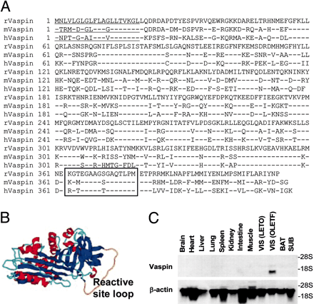
Amino acid sequence,
structural analyses, and gene expresion of vaspin in various
tissues. (A) Amino acid sequence of rat, mouse, and
human vaspins. Signal peptides are underlined, and reactive
site loop is boxed. (B) Automated protein structure
homology modeling by SWISS-MODEL predicted the presence of
three -sheets
(blue), nine -helices
(red), and one reactive site loop (yellow). (C) Northern
blot analyses of vaspin in various organs of obese 30-wk-old
OLETF and visceral adipose tissue of lean 6-wk-old LETO rats.
A single transcript is observed in visceral (VIS) fat of OLETF
rats. BAT, brown adipose tissue. Hida K, et al. Proc Natl
Acad Sci U S A. 2005 Jul 26;102(30):10610-5
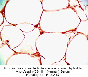
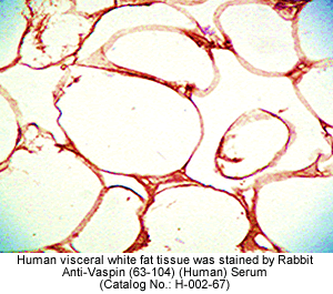
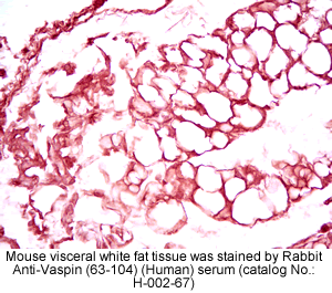
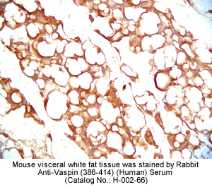
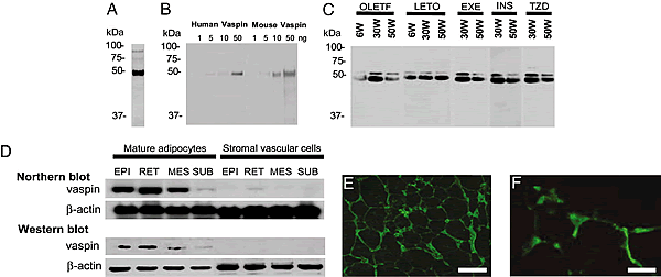
Expression
of vaspin in 293T cells, adipocytes, and stromal vascular
cells. (A) Western blot analysis of mouse vaspin
by using supernatants of 293T cultured cells transfected
with AxCAmOL64 and polyclonal antivaspin Ab. A prominent
band of 45
kDa is observed. (B) Western blots of purified
recombinant human and mouse vaspins, derived from E.
coli by using pET expression system. (C) Western
blot analyses of vaspin by using sera of OLETF and LETO
rats, and OLETF rats with EXE, OLETF rats administered TZD
and insulin (INS). A major 45-kDa
band and a minor 50-kDa
band are seen at 30 wk. Band intensity is notably less at
50 wk in OLTEF rats, but it seems to be normalized with
insulin and TZD treatments. (D) Expression of vaspin
in mature adipocytes and stromal vascular cells isolated
from epidydimal (EPI), retroperitoneal (RET), MES, and SUB
WATs as analyzed by Northern and Western blot analyses.
Expression is confined to adipocytes of visceral WATs and
not to stromal endothelial or vascular cells. (E
and F) Expression in the adipocytes was confirmed
by immunofluorescence microscopy in 30-wk-old OLETF rats.
(Scale bars: E, 100 µm; F, 25 µm.). Hida
K, et al. Proc Natl Acad Sci U S A. 2005 Jul 26;102(30):10610-5
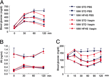
Profiles of glucose
tolerance and insulin sensitization tests after administration
of vaspin and insulin. (A) Glucose tolerance test.
rhVaspin or PBS was i.p. injected into ICR mice fed STD chow
or HFHS chow before glucose administration. Glucose levels
were significantly reduced with the administration of vaspin
(*, P < 0.01). (B) Insulin levels during
the glucose tolerance test were unaltered, and HFHS mice remained
hyperinsulinemic. (C) Insulin tolerance test. rhVaspin
or PBS was i.p. injected into mice before insulin administration.
Blood glucose levels were lowered in the HFHS group receiving
insulin plus vaspin (*, P < 0.05; **, P
< 0.01). All data are mean ± SEM (n = 10). Hida
K, et al. Proc Natl Acad Sci U S A. 2005 Jul 26;102(30):10610-5
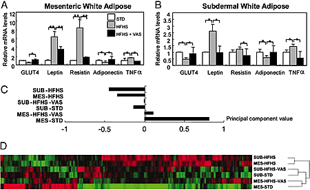
Effect of vaspin on
leptin, resistin, TNF,
GLUT4, and adiponectin in obese ICR mice fed with HFHS chow.
Vaspin administration reverses gene expression profile of
WATs. (A and B) Injection of rhVaspin suppressed
gene expression of leptin, resistin, and TNF
and increased the expression of glucose transporter-4 and
adiponectin in obese ICR mice with HFHS chow. Data are mean
± SEM (n = 5). *, P < 0.05; **, P
< 0.01. (C) Principal component analysis showing
that the HFHS-chow-induced gene expression profile in MES
and SUB fats is distinct from that of ICR mice with STD chow
and after injection of rhVaspin (VAS). (D) Hierarchical
clustering analysis seems to divide in two distinct groups,
HFHS and STD chow groups. Both the MES and the SUB gene expression
profile in rhVaspin-treated ICR mice fed with HFHS chow is
on the same branch as mice fed STD chow. Hida K, et al. Proc
Natl Acad Sci U S A. 2005 Jul 26;102(30):10610-5