| ProSAAS-derived
Peptides 
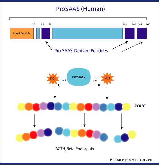
ProSAAS is a newly discovered protein with a neuroendocrine
distribution generally similar to that of prohormone convertase
1 (PC1), a peptide processing endopeptidase. ProSAAS mRNA
is broadly distributed among neurons in brain and other neuroendocrine
tissues (pituitary, adrenal, pancreas).
Overexpression of ProSAAS in the AtT-20 cells substantially
reduces the rate of processing of POMC (endogenous prohormone
proopiomelanocortin) [1]. In wild type mouse brain and pituitary,
the majority of ProSAAS is processed into smaller peptides
including Little SAAS, PEN and big LEN. [2] A few obesity
related propeptides have been processed by PC1 and obesity
has been observed to be associated with mutations in the human
PC1 gene[3]. Does proSAAS-derived peptides play
a role in the development of obesity?
1. Fricker, L.D., et al. J Neurosci 20,639-648(2000)
2. Mazhavia N., et al. ProSAAS processing in mouse brain
and pituitary. JBC Nov. 27, 2000
3. Jackson, R.S., et al. Nat Gene 16,303-306(1997)

|
| Schematic representation of the PC1-specific inhibitor
proSAAS. The presence of two PC-like cleavage sites
in proSAAS and its site of interaction with the catalytic
domain of PC1 are illustrated. The selected proSAAS-(235-246)
peptide used in the current study is also indicated.
hproSAAS, human proSAAS. Basak A, et al. J Biol Chem.
2001 Aug 31;276(35):32720-8 |
|
| Dixon plots for inhibition of mPC1 by proSAAS-(235-246)
(A) and proSAAS-(235-244) (B). The graphical plots
were obtained with data using mPC1 (5 µl) and pERTKR-MCA
as substrate at concentrations 12.5 (S1), 25 (S2),
50 (S3), and 100 µM (S4). The inhibition study was
conducted with a 15-min preincubation between the
enzyme and the inhibitor, following which the substrate
pERTKT-MCA was added. The fluorescence readings were
measured after a 6-h reaction at 37 °C (see "Experimental
Procedures"). hproSAAS, human proSAAS. Basak A, et
al. J Biol Chem. 2001 Aug 31;276(35):32720-8 |
|
| Effect of synthetic proSAAS C-terminal
peptides on the activity of purified PC1. The peptides
were tested with purified PC1 (63 ng) and 100 µM substrate,
as described under "Materials and Methods." The data
shown were obtained after 90 min of incubation at
25 °C. The experiment was performed three times with
less than 10% variation. Qian Y, et al. J Biol Chem.
2000 Aug 4;275(31):23596-601 |
|
| Time-dependent inhibition of mPC1 activity by proSAAS-(235-244)
(192 nM). mPC1 (5 µl, containing 3.9 nmol of AMC released/h)
was preincubated separately with 192 nM of proSAAS-(235-244)
for 0, 5, 15, and 30 min. Following each preincubation
period, pERTKR-MCA (100 µM) was added, and the progress
curves were followed by fluorescence for 30 min. The
enzyme inhibition was determined by the initial slope
of various progress curves. Basak A, et al. J Biol
Chem. 2001 Aug 31;276(35):32720-8 |
|
| Progress curves over a period of 1,000 s showing
inhibition of PC1, furin, PACE4, and PC7 activity
in the presence of varying concentrations of proSAAS-(235-244).
The enzyme activity was measured against the fluorogenic
substrate pERTKR-MCA (100 µM) in the presence of increasing
concentrations of proSAAS-(235-244) (as indicated
in the right side of each panel). The release of fluorescence
was followed over a period of 10 min. The inset in
the PC7 panel is the progress curve monitored over
a period of 1 h. Basak A, et al. J Biol Chem. 2001
Aug 31;276(35):32720-8 |
|
| Northern blot analysis of proSAAS mRNA in human
tissues. Northern blots containing poly(A) RNA (Clontech)
were probed with 32P-labeled human proSAAS cDNA, as
described in Materials and Methods, and were exposed
to x-ray film for 2 hr at 80°C. Fricker LD, et al.
J Neurosci. 2000 Jan 15;20(2):639-48 |
| |
| |
|
| Deletion mutational analysis of the PC1 inhibitory
region of proSAAS. The proSAAS-derived peptides previously
identified in Cpefat/Cpefat mouse tissues and in AtT-20
cells are indicated by shading; the names of several
of these peptides are indicated on the top line and
refer to internal amino acid sequences (KEP, SAAS,
PEN, and LEN). Single (R) and dibasic (RR or KR) processing
sites are indicated. Fusion constructs consisting
of GST with C-terminal extensions of the indicated
portions of rat proSAAS were created in the pGEX-2T
vector (Amersham Pharmacia Biotech) as described under
"Materials and Methods." Protein was expressed in
bacteria and purified on glutathione-agarose. The
amount of protein was quantitated from the absorption
at 280 nm, using the calculated extinction coefficient
for each construct. The construct 231-246 was created
as both a GST fusion protein and as a synthetic peptide,
and the constructs 221-242, 245-260, 245-254, 34-59,
34-40, and 42-59 were created only as synthetic peptides.
The PC1 assay was performed with 5 µM substrate, using
media from PC1-expressing baculovirus as described
under "Materials and Methods." All constructs were
tested at 1 µM in at least two separate experiments,
with similar results. Yes indicates greater than 50%
inhibition, whereas no indicates less than 10% inhibition;
none of the constructs showedpartial inhibition. Qian
Y, et al. J Biol Chem. 2000 Aug 4;275(31):23596-601 |
|
Amino acid identity among human, rat, and mouse proSAAS.
Asterisks below the sequence denote residues conserved
in all three species. Open arrowhead, Signal peptide
cleavage site. Double arrows, Paired basic cleavage
sites (RR, KR) that are used in the Cpefat/Cpefat mouse.
Single arrows, KxxR cleavage site used in the Cpefat/Cpefat
mouse. Half arrows, Additional predicted cleavage sites.
The sequences of the peptides that correspond to those
found in the Cpefat/Cpefat mice (but without the C-terminal
basic residues that would be removed by active CPE)
are indicated by lines. Fricker LD, et al. J Neurosci.
2000 Jan 15;20(2):639-48
|
|


