|
|
|
Endothelin,
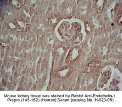
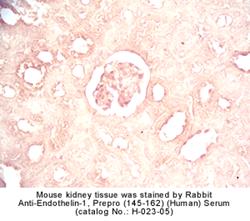
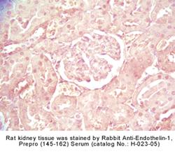
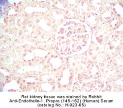
Endothelin-like Peptides & Prepro C-Terminal Peptide |
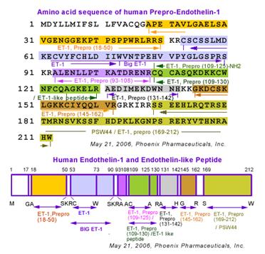
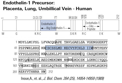
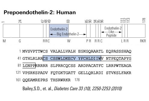
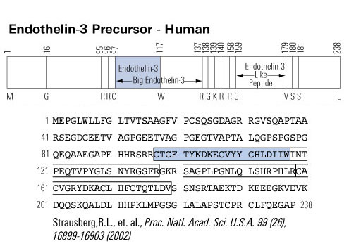
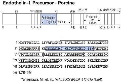
Endothelins are a family of potent
vasoconstrictor peptides comprising three isoforms. It is found in a host of
mammalian species and is thought to have a potent effect in both the cardiovascular
and central nervous systems. Endothelin-1 (ET-1), Endothelin-2 (ET-2) and
Endothelin-3 (ET-3) have 21 amino acids and two disulfide bridges. Endothelin
is found to circulate in the blood in significant levels and is thought to be
involved with increasing blood pressure upon receptor binding
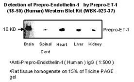
Big Endothelins are
the precursors to Endothelins. They are cleaved by Endothelin Converting Enzyme (ECE) to the mature 21 amino acid peptide.
Steinhelper M.E., Circulation Research 72, No. 5,
984 (1993)
Maack, T., Annu. Rev. Physiol. 54, 11 (1992)
Brenner, B.M. et al., Physiol. Rev. 70, 665 (1990)
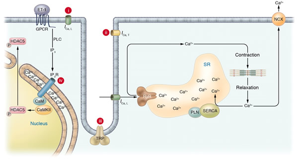
Schematic of potential Ca2+ sources that might be specialized to regulate reactive signaling pathways in cardiac myocytes. (i) Some L-type Ca2+ channels (ICa,L) are not associated with the junctional complex and hence could be involved in providing a local Ca2+ signal in specific membrane-associated compartments. (ii) T-type Ca2+ channels (ICa,T) are reexpressed in hypertrophic states where they could provide Ca2+ in specific microenvironments associated with the sarcolemma to affect reactive signaling pathways. (iii) Capacitative or store-operated Ca2+ entry through transient receptor potential (TRP) channels, alone or in conjunction with (iv) InsP3R-mediated release of Ca2+ from the ER/nuclear envelope, could also provide a highly localized Ca2+ pool for controlling reactive signaling pathways in cardiac myocytes. Signaling from G protein–coupled receptors (GPCRs) activates PLC and generates InsP3, causing a perinuclear Ca2+ signal through the InsP3R, resulting in CamKII activation and HDAC5 nuclear export, as proposed by Wu et al. (2). IP3, InsP3; IP3R, InsP3R; PLN, phospholamban; NCX, Na+/Ca2+ exchanger; RyR, ryanodine receptor; CaM, calmodulin; SERCA, SR/ER Ca2+-ATPase; P, phosphate.
Molkentin, J. D. J. Clin. Invest. 2006;116:623-626
| Intracellular Ca2+ cycling and associated signaling pathway in cardiomyocytes. On a beat-by-beat basis, a calcium transient is evoked by the initial influx of a small amount of Ca2+ through the LTCC and the subsequent large-scale Ca2+ release from the SR through the RyR. During diastole, cytosolic Ca2+ is taken up into the SR by the PLN-regulated SERCA2a pump. ß receptor–mediated PKA stimulation regulates this Ca2+ cycling by phosphorylating LTCC, RyR, and PLN. In normal hearts, sympathetic stimulation activates ß1-adrenergic receptor, which in turn stimulates the production of cAMP by adenylyl cyclase and thereby activates PKA. PKA phosphorylates PLN and RyR, both of which contribute to an increased intracellular Ca2+ transient and enhanced cellular contractility (pink zone signal). PP1 and PP2A regulate the dephosphorylation process of these Ca2+ regulatory proteins (RyR, PLN, LTCC) (blue zone signaling). Activation of the Gq-coupled receptors (angiotensin II receptor, endothelin 1 receptor, or -adrenergic receptor) activates PLC, which in turn activates PKC-. The PKC- phosphorylates I-1, augmenting the activity of PP1 and causing hypophosphorylation of PLB. The PLB hypophosphorylation inhibits SERCA2a activity, thereby decreasing SR Ca2+ uptake. The increased Ca2+ level in the cytosol activates CAMKII, which affects the functions of RyR and PLN. Activation or deactivation of these molecules at a node in the signaling cascade affects beat-by-beat Ca2+ cycling, and such maneuvers have recently been highlighted as potential new therapeutic strategies against HF. , G protein subunit ; ß, G protein subunit ß; , G protein subunit ; AC, adenylyl cyclase; PLC, phospholipase C. |
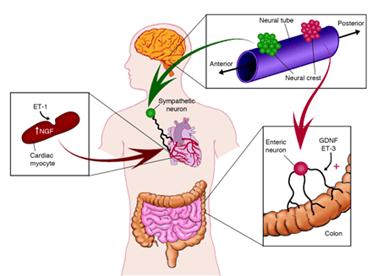
Distinctive pairing of endothelins and neurotrophic factors promotes target innervation during development. Although the sequential action of ET-1 to induce myocyte production of NGF is suggested to promote sympathetic innervation of the heart, more complex interactions of ET-3 and GDNF occur in the patterning of the enteric nervous system.
Barbara L. Hempstead J. Clin. Invest. 113:811-813 (2004)
| 
|
|
() |
() | |
| 
|
|
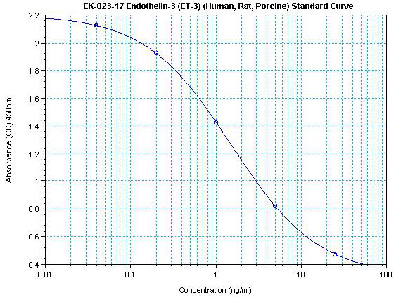
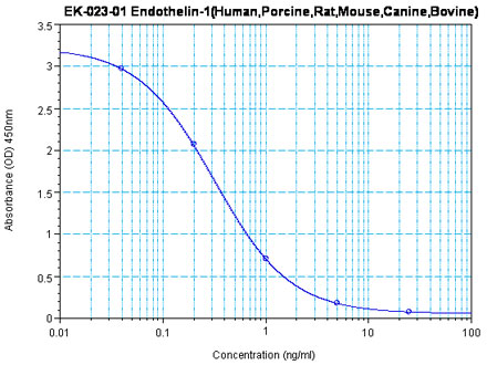
Linear Range: 0.13-1.72 ng/ml
|
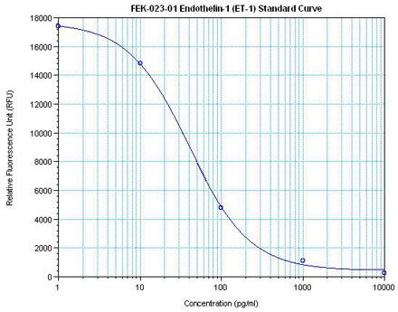
Linear Range: 17.8-185 pg/ml
7 times more sensitive than normal EIA Kits |
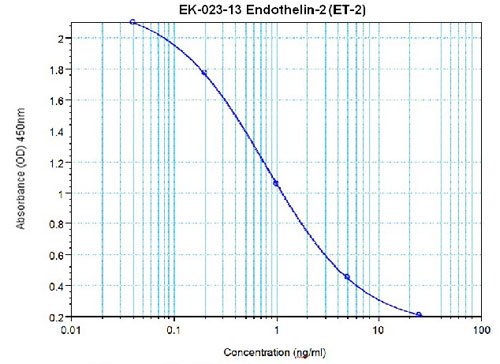
Linear Range: 0.11-1.21 ng/ml
Linear Range 0.33-10.2 ng/ml
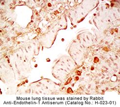
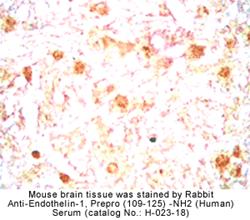
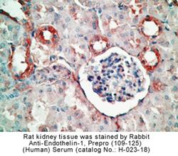
Tips: See More Antibody Stainings, Immunoassay Kits Curves and Sequences by clicking the tabs on the top.
|
|
|
%Endothelin%
|
|
|