We have used rat cDNA microarrays to identify adipocyte-specific genes that could play an important role in adipocyte differentiation or function. Here, we report the cloning and identification of a 2.0-kb mRNA coding for a putative protein that we have designated as desnutrin. The novel gene is expressed predominantly in adipose tissue, and its expression is induced early during 3T3-L1 adipocyte differentiation. Desnutrin mRNA levels were regulated by the nutritional status of animals, being transiently induced during fasting. In vitro desnutrin gene expression was up-regulated by dexamethasone in a dose-dependent manner but not by cAMP, suggesting that glucocorticoids could mediate the increase in desnutrin mRNA levels observed during fasting. Desnutrin mRNA codes for a 486-amino acid putative protein containing a patatin-like domain, characteristic of many plant acyl hydrolases belonging to the patatin family. Confocal microscopy of enhanced green fluorescent protein-tagged desnutrin protein-transfected cells showed that the fusion protein localized in the cytoplasm. Moreover, cells overexpressing desnutrin by transfection showed an increase in triglyceride hydrolysis. Interestingly, we also found that the desnutrin gene expression level was lower in ob/ob and db/db obese mouse models. Overall, our data suggest that the newly identified desnutrin gene codes for an adipocyte protein that may function as a lipase and play a role in the adaptive response to a low energy state, such as fasting, by providing fatty acids to other tissues for oxidation. In addition, decreased expression of desnutrin in obesity models suggests its possible contribution to the pathophysiology of obesity.
Villena JA, et al. J Biol Chem. 2004 Nov 5;279(45):47066-75.
Epub 2004 Aug 27.
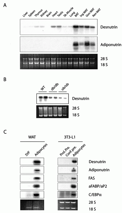
Desnutrin mRNA levels in various adult mouse
tissues and cells. A, 10 µg of total RNA from various mouse
tissues was analyzed by Northern blotting and hybridized with radiolabeled
desnutrin and adiponutrin cDNA probes. Sk, skeletal; Ing, inguinal;
Gon, gonadal; Ren, renal. B, shown is desnutrin mRNA expression
in gonadal WAT from 12-h fasted wild-type (WT), db/db, and ob/ob
mice. C, 5 µg of total RNA from cells of the stromal vascular
fraction (SVF) or adipocytes isolated from mouse inguinal adipose
tissue (left panels) or 10 µg of total RNA from 3T3-L1 cells
at the indicated days of differentiation (right panels) was examined
by Northern blot analysis for the expression of desnutrin and various
adipocyte markers. Prol. Pre., proliferating preadipocytes; Conf.
pre., confluent preadipocytes; FAS, fatty-acid synthase; aFABP,
adipocyte fatty acid-binding protein.
Villena JA, et al. J Biol Chem. 2004 Nov 5;279(45):47066-75.
Epub 2004 Aug 27.
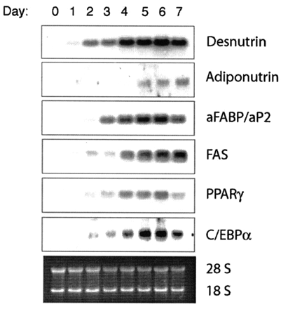
Desnutrin mRNA levels during adipocyte differentiation of 3T3-L1
cells. Two-day post-confluent 3T3-L1 preadipocytes (day 0) were
induced to differentiate by treatment with 1 µM dexamethasone
and 0.5 mM MIX for 2 days and then maintained in differentiation
medium for an additional 5 days. Ten µg of total RNA prepared
from cells collected at the indicated time points was examined for
the expression of desnutrin and other adipocyte markers by Northern
blot analysis. aFABP, adipocyte fatty acid-binding protein; FAS,
fatty-acid synthase; PPAR, peroxisome proliferator-activated receptor-gamma.
Villena JA, et al. J Biol Chem. 2004 Nov 5;279(45):47066-75.
Epub 2004 Aug 27.
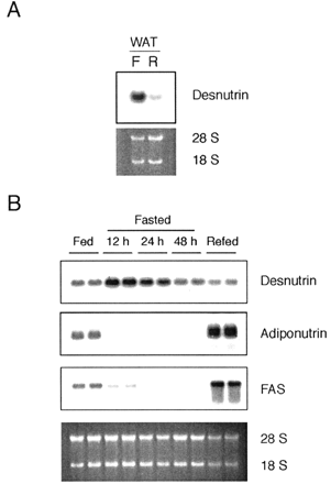
Nutritional regulation of desnutrin mRNA levels. A, expression of
desnutrin mRNA in WAT from mice fasted for 24 h (F) or fasted and
then refed for 12 h (R) as assessed by Northern blot analysis; B,
time course analysis of desnutrin and adiponutrin mRNA expression
levels in WAT during fasting. Mice were fasted for 12, 24, or 48
h, and total RNA was extracted for examination of desnutrin mRNA
levels by Northern blot analysis. Desnutrin mRNA levels in WAT were
compared with those in mice that were fasted for 48 h and subsequently
fed for 12 h. FAS, fatty-acid synthase.
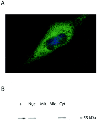
Subcellular localization of desnutrin-EGFP
fusion protein. A, COS-7 cells were transfected with the desnutrin-EGFP
expression vector, and localization of the fusion protein was assessed
by confocal microscopy. B, COS-7 cells were transfected with an
HA-tagged desnutrin expression vector, and nuclear (Nuc.), mitochondrial
(Mit.), microsomal (Mic.), and cytosolic (Cyt.) fractions were prepared
as described under "Experimental Procedures." Five µg
of protein from each fraction was subjected to SDS-PAGE, transferred
to a polyvinylidene difluoride membrane, and analyzed for the presence
of HA-desnutrin using anti-HA antibody. As a positive control for
the immunodetection, 10 µg of whole cell lysate was used.
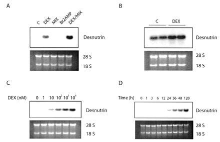
Regulation of desnutrin mRNA levels by glucocorticoids. A, confluent
preadipocytes treated for 48 h with 1 µM dexamethasone (DEX),
0.5 mM MIX, 0.5 mM dibutyryl cAMP (Bt2AMP), or 1 µM dexamethasone
and 0.5 mM MIX for 48 h (DEX/MIX). After the incubation period,
cells were harvested, and desnutrin mRNA levels were analyzed by
Northern blotting. B, desnutrin mRNA levels in fully differentiated
adipocytes untreated (C) or treated for 48 h with 1 µM dexamethasone.
C, dose-dependent induction of desnutrin mRNA by dexamethasone in
3T3-L1 preadipocytes. D, time course analysis of desnutrin mRNA
expression during dexamethasone treatment in 3T3-L1 preadipocytes.
Cells were treated with 1 µM dexamethasone for the indicated
times.
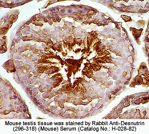
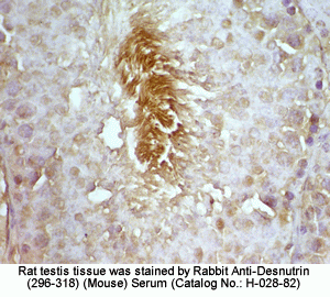
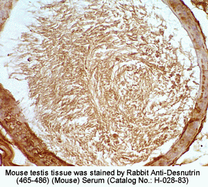
Mapping in Mouse & Rat Testis Tissue by Desnutrin (465-486) (Mouse) - Antibody (H-028-83)
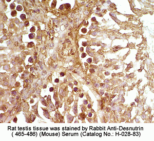
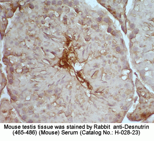
Mapping in Mouse & Rat Testis Tissue by Desnutrin (296-318) (Mouse) - Antibody (H-028-82)
