A Bioactive Lipid-derived Factor Inhibiting Food Intake by Activation of PPAR-alpha & GPR119
FOOD INTAKE REGULATES OLEOYLETHANOLAMIDE FORMATION AND DEGRADATION IN THE PROXIMAL SMALL INTESTINE
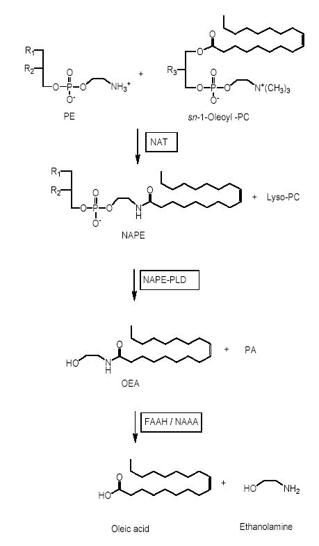
Oleoylethanolamide (OEA) is a lipid mediator that inhibits food intake by activating the nuclear receptor peroxisome proliferator-activated receptor- α (PPAR-α). In the rodent small intestine, OEA levels decrease during food deprivation and increase upon refeeding, suggesting that endogenous OEA may participate in the regulation of satiety. Here we show that feeding stimulates OEA mobilization in the mucosal layer of rat duodenum and jejunum, but not in the serosal layer from the same intestinal segments, in other sections of the gastrointestinal tract (stomach, ileum, colon), or in a broad series of internal organs and tissues (e.g., liver, brain, heart, plasma). Feeding also increases the levels of other unsaturated fatty-acid ethanolamides (FAEs) (e.g., linoleoylethanolamide) without affecting those of saturated FAEs (e.g., palmitoylethanolamide). Feeding-induced OEA mobilization is accompanied by enhanced accumulation of OEA-generating N-acyl phosphatidylethanolamines (NAPEs), increased activity and expression of the OEA-synthesizing enzyme NAPE-phospholipase D (NAPE-PLD), and decreased activity and expression of the OEA-degrading enzyme fatty-acid amide hydrolase (FAAH). Immunostaining studies revealed that NAPE-PLD and FAAH are expressed in intestinal enterocytes and lamina propria cells. Collectively, these results indicate that nutrient availability controls OEA mobilization in the mucosa of the proximal intestine through a concerted regulation of OEA biosynthesis and degradation.
Fu J. et al. J Biol Chem. 2007 January 12; 282(2): 1518–1528.
Expression and distribution of Gpr119 in the pancreatic islets of mice and rats : Predominant localization in pancreatic polypeptide-secreting PP-cells.
The GPR119 was recently shown to be activated by oleoylethanolamide (OEA), a naturally occurring bioactive lipid with hypophagic and anti-obesity effects. In this study, we have cloned and characterized its murine counterpart, Gpr119. The full-length cDNA contained an open reading frame of 1008 bp encoding a 335-amino acid protein. The genomic organization of Gpr119 was unique, having a 3'-untranslated second exon that was also involved in an alternative splicing event. Gene expression analyses confirmed its specific expressions in pancreatic islets and two endocrine cell-lines, MIN6 and αTC1. Immunohistochemistry and double-immunofluorescence studies using a specific antibody revealed the predominant Gpr119 localization in pancreatic polypeptide (PP)-cells of islets. No definitive evidence of Gpr119-immunoreactivity in adult β- or α-cells was obtained. The Gpr119 mRNA levels were elevated in islets of obese hyperglycemic db/db mice as compared to control islets, suggesting a possible involvement of this receptor in the development of obesity and diabetes.
SAKAMOTO Yukiko et al. Biochemical and biophysical research communications. 2006, vol. 351, no2, pp. 474-480 [7 page(s) (article)]
GPR119, a novel G protein-coupled receptor target for the treatment of type 2 diabetes and obesity
GPR119 is a G protein-coupled receptor expressed predominantly in the pancreas (beta-cells) and gastrointestinal tract (enteroendocrine cells) in humans. De-orphanization of GPR119 has revealed two classes of possible endogenous ligands, viz., phospholipids and fatty acid amides. Of these, oleoylethanolamide (OEA) is one of the most active ligands tested so far. This fatty acid ethanolamide is of particular interest because of its known effects of reducing food intake and body weight gain when administered to rodents. Agonists at the GPR119 receptor cause an increase in intracellular cAMP levels via G(alphas) coupling to adenylate cyclase. In vitro studies have indicated a role for GPR119 in the modulation of insulin release by pancreatic beta-cells and of GLP-1 secretion by gut enteroendocrine cells. The effects of GPR119 agonists in animal models of diabetes and obesity are reviewed, and the potential value of such compounds in future therapies for these conditions is discussed.
Overton HA, Fyfe MC, Reynet C. Br J Pharmacol. 2008 Mar;153 Suppl 1:S76-81. Epub 2007 Nov 26.
Fu J. et al. J Biol Chem. 2007 January 12; 282(2): 1518–1528.
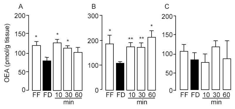
Feeding-induced OEA mobilization in the rat proximal small intestine.
Effect of free-feeding (FF), 24-h food deprivation (FD) and refeeding (10, 30 and 60 min) on OEA levels in duodenum (A), jejunum (B) and ileum (C).*p < 0.05, ** p < 0.01 vs FD, n = 10–12.
Fu J. et al. J Biol Chem. 2007 January 12; 282(2): 1518–1528.
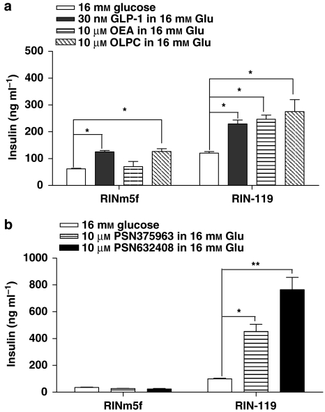
GPR119 agonists increase insulin secretion in RIN cell lines. (a and b) The RINm5f
and RIN-119 cells were stimulated by 16mM glucose,
with or without the presence of 30nM glucagon-like peptide-1, 10μM oleoylethanolamide, 10μM oleoyl-lysophosphatidylcholine,
10μM PSN375963 and PSN632408 as indicated. The
insulin levels from the triplicates were measured for each treatment and data were
presented as ng insulin per ml (mean±s.e.mean).
*P<0.05; **P<0.01 using
ANOVA-Bonferroni, compared with insulin induced by 16mM glucose alone in the same cell line.
Br J Pharmacol. Published online 2008 August 25. doi: 10.1038/bjp.2008.337.
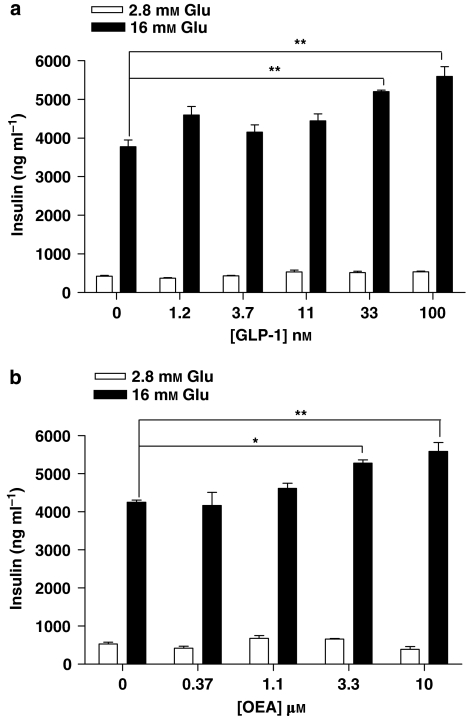
Glucagon-like peptide-1 (GLP-1) and oleoylethanolamide (OEA) increase
glucose-stimulated insulin secretion.
MIN6c4 cells were stimulated with the
indicated concentrations of (a) GLP-1, (b) OEA, in the presence of 2.8mM
glucose or 16mM glucose.
The insulin levels were measured after 2h incubation.
Three replicates were measured for every treatment and data presented as ng
insulin ml−1 (mean±s.e.).
*P<0.05; **P<0.01 using ANOVA-Bonferroni,
compared with insulin induced by glucose alone.