a Novel skeletal muscle-derived and Bone-specific Secreted Protein that Modulates the Osteoblast Phenotype
Musclin, a novel skeletal muscle-derived secretory factor
Skeletal muscle is involved in the homeostasis of
glucose and lipid metabolism. We hypothesized that the skeletal
muscle produces and secretes bioactive factor(s), similar to
adipocytokines secreted by fat tissue. Here, we report the
identification of a novel secretory factor, musclin, by signal
sequence trap (SST) of mouse skeletal muscle cDNAs. Musclin cDNA
encoded 130 amino acids, including N-terminal 30-amino acid signal
sequence. Musclin protein contained a region homologous to
natriuretic peptide (NP) family, and KKKR, a putative serine
protease cleavage site, similar to NP family. Full-length musclin
protein and KKKR-dependent cleaved form were secreted in media of
musclin cDNA-transfected mammalian cell cultures. Musclin mRNA was
expressed almost exclusively in the skeletal muscle of mice and
rats. Musclin mRNA levels in skeletal muscle were markedly low in
fasted, increased upon re-feeding, and were low in
streptozotocin-treated insulin-deficient mice. Musclin mRNA
expression was induced at late stage in the differentiation of C2C12
myocytes. In myocytes, insulin increased while epinephrine,
isoproterenol and forskolin reduced musclin mRNA, all of which are
known to increase the cellular content of cyclic AMP, a counter
regulator to insulin. Pathologically, overexpression of musclin mRNA
was noted in the muscles of obese insulin-resistant KKAy and db/db
mice. Functionally, recombinant musclin significantly attenuated
insulin-stimulated glucose uptake in myocytes. In conclusion, we
identified musclin, a novel skeletal muscle-derived secretory
factor. Musclin expression level is tightly regulated by nutritional
changes and its physiological role could be linked to glucose
metabolism.
Nishizawa H, et al. J Biol Chem. 2004 Mar 24 [Epub ahead of print]
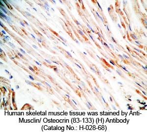
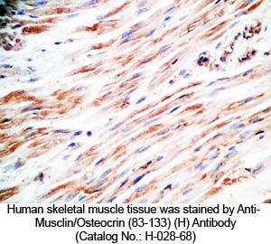
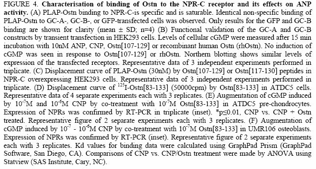
Osteocrin is a specific ligand of the natriuretic peptide clearance receptor that modulates bone growth
Osteocrin (Ostn) is a recently discovered secreted protein,
produced by cells of the osteoblast lineage, which shows a well
conserved homology with members of the natriuretic peptide (NP)
family. We hypothesized that Ostn could interact with the
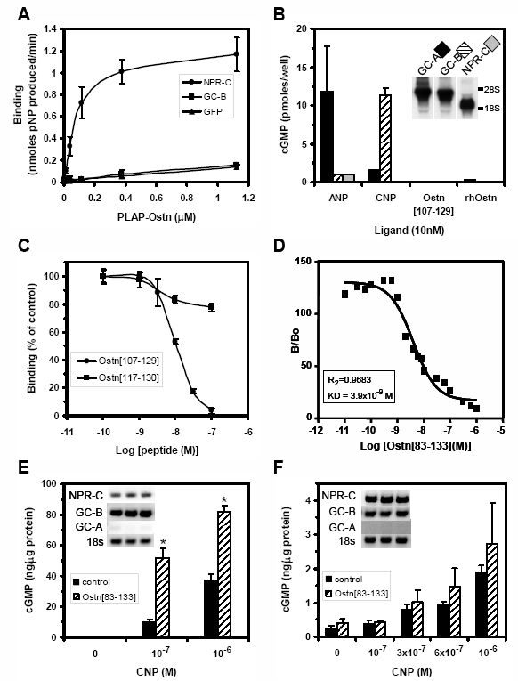
NP-receptors thereby modulating NP actions on the skeleton. Ostn
binds specifically and saturably to the NPR-C receptor with a Kd
~5nM with no binding to the GC-A or GC-B receptors. Deletion of
several of the residues deemed important for NP binding to NPR-C
lead to abolition of Ostn binding confirming the presence of a
"natriuretic motif". Functionally, Ostn was able to augment
CNP-stimulated cGMP production in both pre-chondrocytic (ATDC5) and
osteoblastic (UMR106) cells suggesting increased NP levels due to
attenuation of NPR-C associated NP-clearance. Ostn-transgenic mice
displayed elongated bones and a marked kyphosis associated with
elevated bone cGMP levels suggesting elevated natriuretic peptide
activity contributed to the increased bone length possibly through
an increase in growth plate chondrocyte proliferation. Thus we have
demonstrated that Ostn is a naturally occurring ligand of the NPR-C
clearance receptor and may act to locally modulate the actions of
the natriuretic system in bone by blocking the clearance action of
NPR-C thus locally elevating levels of CNP.
Moffatt P, et al. J Biol Chem. 2007 Oct 19; [Epub ahead of print]
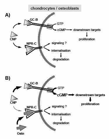
Ostn blocks the clearance action of NPR-C thereby increasing
activity of the NPs in the
bone compartment (A) Both the GC-B and
NPR-C receptors are expressed in osteoblasts and
chondrocytes,
thus the activity of the NPs (normally CNP in the skeleton) is
governed by the distribution of CNP between the signal mediating
GC-B receptor and the NPR-C clearance receptor. (B) In the presence
of Ostn, CNP access to NPR-C is blocked leading to increased binding
of CNP to the GC-B receptor. This results in an increase in
GC-B-mediated cGMP production, magnifying the downstream biological
effects of the natriuretic system, which in the skeleton leads to
elongated bones.
Moffatt P, et al. J Biol Chem. 2007 Oct 19; [Epub ahead of print]
Predicted osteocrin protein structure and secretion.
A, predicted amino acid sequences from human, bovine, mouse, rat, chicken, and snake (partial) cDNAs (see
"Materials and Methods"). The two dibasic cleavage sites, 76KKKR79
and 110KKR112, are well conserved (boxed). Shaded
residues represent non-conserved residues from the human sequence.
Note that the C-terminal half of the protein is highly conserved. B, immunofluorescent localization with a human
osteocrin-specific antibody demonstrates overexpressed osteocrin is
clearly visible in the secretory apparatus of HEK293A fibroblasts
and UMR106 osteosarcoma cells (x400) colocalizing with the 58K Golgi
protein-specific antibody. C, the majority of osteocrin is
detected in the medium of HEK293 cells by Western blot when
transiently transfected (lanes 1 and 2). Residual
protein in the secretory apparatus is detected in the cell lysate
(lanes 4 and 5). Two exposures of the Western blot
are shown to illustrate the underrepresented smaller processed
fragments, with the lower panel representing a longer
exposure. A doublet can be seen at the expected full-length size of
11.4 kDa and smaller processed fragments at ~ 5 kDa (lane
1). Mutation of the KKKR site to AS abolishes processing of the
protein leaving only full-length 11.4 kDa bands (lane 2).
Equal proportions of media or cell lysate from cultured cells were
loaded. WT, wild type; Mut, mutated.
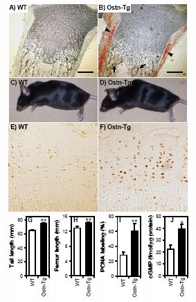
Ostn-transgenic (Ostn-TG) mice have longer bones.
Ostn-TG mice
with over-expression of Ostn in osteoblast lineage-specific cells
were generated using the 3.6kb collagen type I
promoter. Immunohistochemistry using an Ostn-specific antibody on
4-day-old tibia of wild type (WT)
(A) and Ostn-TG (B) mice. Ostn
expression in WT is non detectable (A) whereas overexpression in
Ostn-TG is evident (red staining) on cuboidal osteoblasts
adjacent to trabecular bone of the primary spongiosa (arrows),
and within the perichondrium (arrowheads) (B). Counter stain is
methylgreen. Scale bar is 200μm. (C, D) Gross appearance of WT
(C) and Ostn-TG (D) which have a marked kyphosis presumably due to
vertebral overgrowth. (E-F) Proliferation in growth plate
chondrocytes was measured in tibiae of 7- week old male mice using
PCNA immunohistochemistry. Proliferating chondrocytes are clearly
more abundant in Ostn-TG (F) mice than WT littermates (E). Tail (G)
and femur (H) lengths of 8-week old Ostn-TG line 650 males are
significantly longer than wildtype littermates (n=7-12). (I)
Quantification of PCNA immunohistochemistry showed an 80% increase
in staining of growth plate chondrocytes (n=4) (J). cGMP levels were
significantly higher in 10-14 day-old Ostn-TG mice femurs and tibiae
than their WT littermates (n=11-28). *p<0.05; **p<0.01,
Ostn-TG vs. WT. Data are expressed as mean ± SE. Comparison were
made by ANOVA using Statview.
Moffatt P, et al. J Biol Chem. 2007 Oct 19; [Epub ahead of print]
Osteocrin is expressed in osteoblasts.
A, in situ localization of osteocrin transcripts in e16.5 mouse
ribs. Osteocrin is localized within a subset of Cbfa-1-positive
cells in a subperiosteal layer (per). Active mature
osteoblasts are signified by an arrowhead (x200).
B, expression of osteocrin in whole e17.5 fore limb.
Osteocrin is clearly localized in the osteoblasts on the periphery
of the bone (x100). C, in e16.5 mouse tibiae, osteocrin
co-localizes with a subset of Cbfa-1-positive cells, exclusive from
the more mature osteocalcin-positive cells found on the bone surface
and in the trabecular bone (arrowheads) (x400).
Osteocrin is a bone-specific gene.
A, Northern blot showing bone specificity of mouse osteocrin expression. Osteocrin is only detected in neonate and adult long
bones and calvaria. No message was detected in embryonic (e) or adult (ad) non-bone tissues. Five µg total
RNA was loaded. Br, brain; Go, gonads; Te, testes; He, heart; In, intestine;
Ki, kidney; Li, liver; Lu, lung; Sp, spleen. B, osteocrin in detected by Northern
blot in rat spleen. The transcript size of adult rat spleen osteocrin is smaller than that for adult rat calvaria. Fifteen µg of
total RNA was loaded. The position of the 18 S ribosomal RNA band is marked. C, osteocrin expression was detected in embryonic
calvariae and UMR106 cells by RT-PCR. No expression was detected in MG-63, SaoS-2, undifferentiated (-), or differentiated (+) MC3T3.E1
cells. D, Northern blot showing osteocrin expression in femora and calvariae in embryonic (e21), newborn
(p4), growing (1 month, 1m), adult (3 month, 3m), and aged (8-month, 8m) rats. In femora,
osteocrin expression was highest in newborn rats decreasing
significantly in aged rats. Osteocrin expression peaks at 1-month in
calvaria with a less marked decrease in older rats. Twenty-five µg
of total RNA was loaded. ALP, alkaline phosphatase. For
Figs. 4, A-D, GAPDH represents a loading control.
E, immunohistochemistry with an osteocrin-specific antibody
showing localization of osteocrin protein to active osteoblasts
(Ob) in adult mouse tibia. The protein is absent from the
mature osteocytes (Oc). No staining is visible in the
sections stained with pre-immune serum (x400).
Osteocrin is expressed during osteoblast matrix production
and maturation.
Northern blot of a time course of calvarial
primary osteoblast differentiation at confluence and 5 and 10 days
post-confluence in the presence and absence of 10 mM GP (PO4). Osteocrin is expressed
at 5 and 10 days post-confluence but not at confluence. Cultures
maintained in 10 mM GP until 10 days post-confluence exhibit a marked
down-regulation in osteocrin expression. Osteocalcin and osteopontin
expression are highest at 10 days post-confluence and are increased
by GP treatment.
Twenty µg of total RNA was loaded. GAPDH represents a loading
control. Duplicate samples are shown.GP (PO4). Osteocrin is
expressed at 5 and 10 days post-confluence but not at confluence.
Cultures maintained in 10 mM GP until 10 days post-confluence
exhibit a marked down-regulation in osteocrin expression.
Osteocalcin and osteopontin expression are highest at 10 days
post-confluence and are increased by GP treatment. Twenty µg of total
RNA was loaded. GAPDH represents a loading control. Duplicate
samples are shown.
Osteocrin regulates the osteoblast phenotype.
Primary calvarial osteoblastic cultures were treated with
conditioned media from osteocrin or empty vector (control)
transfected HEK293A cells from day 2 to 10 days post-confluence.
A, 45Ca uptake was reduced 60% by osteocrin
conditioned-media. Data is expressed as the mean ± S.E. of the %
45Ca incorporated into the cultures. *, p < 0.01. B, Northern blot demonstrating total repression of
osteocalcin and a marked reduction in alkaline phosphatase
expression in osteocrin treated cultures. Fifteen µg of total RNA
was loaded. GAPDH represents a loading control. Duplicate samples
are shown.